Accelerate Your Biomolecule
and Bioassay Research
and Bioassay Research
sales@neoscientific.com
+1-888.733.6849
+1-617.299.7367 (Int’l)
+1-617.299.7367 (Int’l)
+1-888.733.6849
+1-617.299.7367 (Int’l)
+1-617.299.7367 (Int’l)
For quotations, please use our online quotation form, and you may also contact us by
sales@neoscientific.com
+1-888.733.6849
+1-617.299.7367 (Int’l)
+1-888.733.6849
+1-617.299.7367 (Int’l)
| Reactivity | Human Mouse Rat |
| Tested applications | WB IHC IF |
| Recommended Dilution | WB 1:500 - 1:2000 IHC 1:50 - 1:200 IF 1:50 - 1:200 |
| Calculated MW | 34kDa |
| Observed MW | Refer to Figures |
| Immunogen | Recombinant protein of human AIMP1 |
| Storage Buffer | Store at -20℃. Avoid freeze / thaw cycles. Buffer: PBS with 0.02% sodium azide, 50% glycerol, pH7.3. |
| Synonym | p43; HLD3; EMAP2; SCYE1; EMAPII; |
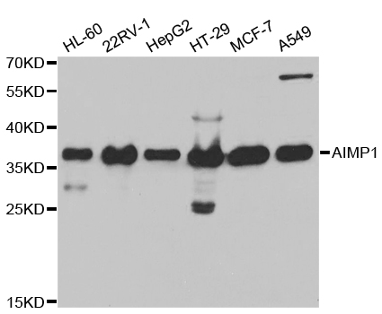
Western blot analysis of extracts of various cell lines, using AIMP1 antibody.
Immunohistochemistry of paraffin-embedded rat spleen using AIMP1 antibody at dilution of 1:100 (x40 lens).
Immunohistochemistry of paraffin-embedded rat kidney using AIMP1 antibody at dilution of 1:100 (x40 lens).

Immunohistochemistry of paraffin-embedded rat heart using AIMP1 antibody at dilution of 1:100 (x40 lens).

Immunohistochemistry of paraffin-embedded human liver cancer using AIMP1 antibody at dilution of 1:100 (x40 lens).

Immunohistochemistry of paraffin-embedded human gastric cancer using AIMP1 antibody at dilution of 1:100 (x40 lens).

Immunohistochemistry of paraffin-embedded mouse kidney using AIMP1 antibody at dilution of 1:100 (x40 lens).

Immunohistochemistry of paraffin-embedded mouse heart using AIMP1 antibody at dilution of 1:100 (x40 lens).

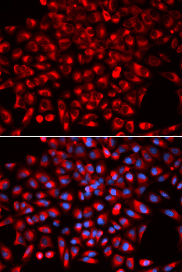
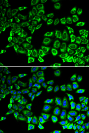
Immunofluorescence analysis of U2OS cell using AIMP1 antibody. Blue: DAPI for nuclear staining.

Immunofluorescence analysis of HeLa cell using AIMP1 antibody. Blue: DAPI for nuclear staining.
The protein encoded by this gene is a cytokine that is specifically induced by apoptosis, and it is involved in the control of angiogenesis, inflammation, and wound healing. The release of this cytokine renders the tumor-associated vasculature sensitive to tumor necrosis factor. The precursor protein is identical to the p43 subunit, which is associated with the multi-tRNA synthetase complex, and it modulates aminoacylation activity of tRNA synthetase in normal cells. This protein is also involved in the stimulation of inflammatory responses after proteolytic cleavage in tumor cells. Multiple transcript variants encoding different isoforms have been found for this gene. A pseudogene has been identified on chromosome 20.
N/A


Copyrights © 2014 NEO Group 245 First Street, 18th Floor, Cambridge MA 02142 888.733.6849


