Accelerate Your Biomolecule
and Bioassay Research
and Bioassay Research
sales@neoscientific.com
+1-888.733.6849
+1-617.299.7367 (Int’l)
+1-617.299.7367 (Int’l)
+1-888.733.6849
+1-617.299.7367 (Int’l)
+1-617.299.7367 (Int’l)
For quotations, please use our online quotation form, and you may also contact us by
sales@neoscientific.com
+1-888.733.6849
+1-617.299.7367 (Int’l)
+1-888.733.6849
+1-617.299.7367 (Int’l)
| Reactivity | Human Mouse Rat |
| Tested applications | WB IHC IF |
| Recommended Dilution | WB 1:500 - 1:2000 IHC 1:50 - 1:100 IF 1:50 - 1:200 |
| Calculated MW | 34kDa |
| Observed MW | Refer to Figures |
| Immunogen | A synthetic peptide of human CDK1 |
| Storage Buffer | Store at -20℃. Avoid freeze / thaw cycles. Buffer: PBS with 0.02% sodium azide, 50% glycerol, pH7.3. |
| Concentration | bd |
| Synonym | CDC2;CDC28A;CDK1;DKFZp686L20222;MGC111195 ; |
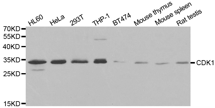
Western blot analysis of extracts of various cell lines, using CDK1 antibody.
Immunohistochemistry of paraffin-embedded human esophageal using CDK1 antibody at dilution of 1:100 (40x lens).
Immunohistochemistry of paraffin-embedded human gastric cancer using CDK1 antibody at dilution of 1:100 (40x lens).

Immunohistochemistry of paraffin-embedded human colon using CDK1 antibody at dilution of 1:100 (40x lens).

Immunohistochemistry of paraffin-embedded human gastric cancer using CDK1 antibody at dilution of 1:100 (40x lens).

Immunohistochemistry of paraffin-embedded mouse spleen using CDK1 antibody at dilution of 1:100 (40x lens).

Immunohistochemistry of paraffin-embedded rat brain using CDK1 antibody at dilution of 1:100 (40x lens).

Immunohistochemistry of paraffin-embedded human lung cancer using CDK1 antibody at dilution of 1:100 (40x lens).

Immunohistochemistry of paraffin-embedded human esophagus using CDK1 antibody at dilution of 1:100 (40x lens).

Immunohistochemistry of paraffin-embedded mouse brain using CDK1 antibody at dilution of 1:100 (40x lens).

Immunohistochemistry of paraffin-embedded human prostate using CDK1 antibody at dilution of 1:100 (x40 lens).

Immunohistochemistry of paraffin-embedded human lung cancer using CDK1 antibody at dilution of 1:100 (x40 lens).

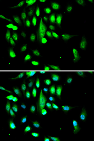
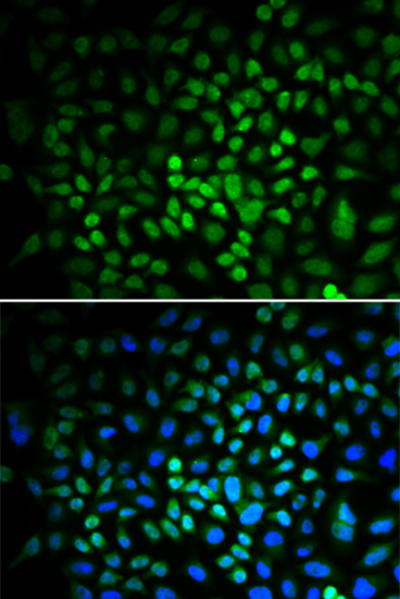
Immunofluorescence analysis of A549 cell using CDK1 antibody. Blue: DAPI for nuclear staining.

Immunofluorescence analysis of HeLa cell using CDK1 antibody. Blue: DAPI for nuclear staining.
The entry of eukaryotic cells into mitosis is regulated by cdc2 kinase activation, a process controlled at several steps including cyclin binding and phosphorylation of cdc2 at Thr161 (1). However, the critical regulatory step in activating cdc2 during progression into mitosis appears to be dephosphorylation of cdc2 at Thr14 and Tyr15 (2). Phosphorylation at Thr14 and Tyr15, resulting in inhibition of cdc2, can be carried out by Wee1 and Myt1 protein kinases (3,4). The cdc25 phosphatase may be responsible for removal of phosphates at Thr14 and Tyr15 and subsequent activation of cdc2 (1,5).
N/A


Copyrights © 2014 NEO Group 245 First Street, 18th Floor, Cambridge MA 02142 888.733.6849


