Accelerate Your Biomolecule
and Bioassay Research
and Bioassay Research
sales@neoscientific.com
+1-888.733.6849
+1-617.299.7367 (Int’l)
+1-617.299.7367 (Int’l)
+1-888.733.6849
+1-617.299.7367 (Int’l)
+1-617.299.7367 (Int’l)
For quotations, please use our online quotation form, and you may also contact us by
sales@neoscientific.com
+1-888.733.6849
+1-617.299.7367 (Int’l)
+1-888.733.6849
+1-617.299.7367 (Int’l)
| Reactivity | Human Mouse Rat |
| Tested applications | WB IHC IF |
| Recommended Dilution | WB 1:500 - 1:2000 IHC 1:50 - 1:200 IF 1:50 - 1:200 |
| Calculated MW | 48kDa |
| Observed MW | Refer to Figures |
| Immunogen | Recombinant protein of human DDB2 |
| Storage Buffer | Store at -20℃. Avoid freeze / thaw cycles. Buffer: PBS with 0.02% sodium azide, 50% glycerol, pH7.3. |
| Concentration | el |
| Synonym | DDBB; FLJ34321; UV-DDB2; |
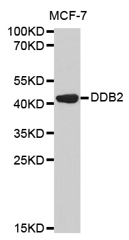
Western blot analysis of extracts of MCF-7 cell line, using DDB2 antibody.
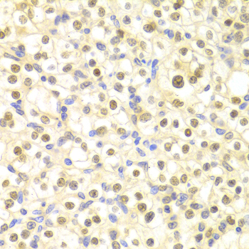
Immunohistochemistry of paraffin-embedded rat heart using DDB2 antibody at dilution of 1:100 (x400 lens).
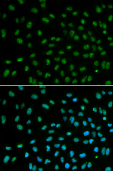
Immunofluorescence analysis of MCF7 cell using DDB2 antibody. Blue: DAPI for nuclear staining.
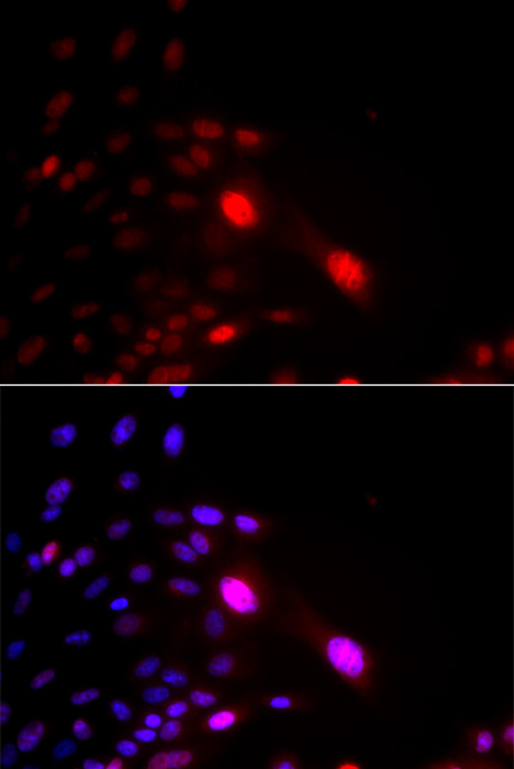
Immunofluorescence analysis of U2OS cell using DDB2 antibody. Blue: DAPI for nuclear staining. DNA damage by a UV-A laser.
Damaged DNA-Binding Protein (DDB) consists of a 127 kDa subunit (DDB-1) and a 48 kDa subunit (DDB-2) that contribute to the formation of the UV-damaged DNA-binding protein complex (UV-DDB) (1-3). In conjunction with CUL4A and ROC-1, the UV-DDB complex forms an E3 ubiquitin ligase that recognizes a broad spectrum of DNA lesions such as cyclobutane pyrimidine dimers, 6-4 photoproducts, apurinic sites and short mismatches. The complex polyubiquitinates components of the nucleotide excision repair pathway (4-6). Loss of DDB activity has been identified in a subset of xeroderma pigmentosum complementation group E (XP-E) patients and has been linked to the deficient repair of cyclobutane pyrimidine dimers in cells derived from these patients (7-10).
N/A


Copyrights © 2014 NEO Group 245 First Street, 18th Floor, Cambridge MA 02142 888.733.6849


