Accelerate Your Biomolecule
and Bioassay Research
and Bioassay Research
sales@neoscientific.com
+1-888.733.6849
+1-617.299.7367 (Int’l)
+1-617.299.7367 (Int’l)
+1-888.733.6849
+1-617.299.7367 (Int’l)
+1-617.299.7367 (Int’l)
For quotations, please use our online quotation form, and you may also contact us by
sales@neoscientific.com
+1-888.733.6849
+1-617.299.7367 (Int’l)
+1-888.733.6849
+1-617.299.7367 (Int’l)
| Reactivity | Human Mouse Rat Other (Wide Range) |
| Tested applications | WB IHC IF IP CHIP CHIPseq |
| Recommended Dilution | WB 1:500 - 1:1000 IHC 1:50 - 1:100 IF 1:50 - 1:200 IP 1:50 - 1:200 ChIP 1:50 - 1:200 CHIPseq 1:50 - 1:200 |
| Calculated MW | 16kDa |
| Observed MW | Refer to Figures |
| Immunogen | A synthetic peptide of human DiMethyl-Histone H3-K79 |
| Storage Buffer | Store at -20℃. Avoid freeze / thaw cycles. Buffer: PBS with 0.02% sodium azide, 50% glycerol, pH7.3. |
| Concentration | qu |
| Synonym | H3K79me2; H3.4; H3/g; H3FT; H3t; MGC126886; MGC126888; |
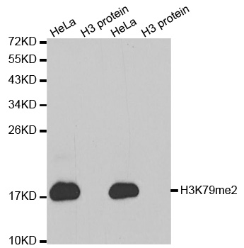
Western blot analysis of extracts of HeLa cell line and H3 protein expressed in E.coli., using H3K79me2 antibody.
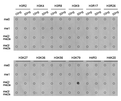
Dot-blot analysis of all sorts of methylation peptides using H3K79me2 antibody.
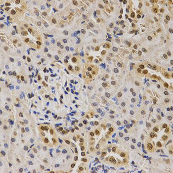
Immunohistochemistry of paraffin-embedded rat kidney tissue using H3K79me2 antibody at dilution of 1:200 (x400 lens).
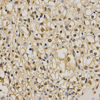
Immunohistochemistry of paraffin-embedded human kidney cancer tissue using H3K79me2 antibody at dilution of 1:200 (x400 lens).

Immunohistochemistry of paraffin-embedded Mouse heart using H3K79me2 antibody at dilution of 1:100 (x400 lens).
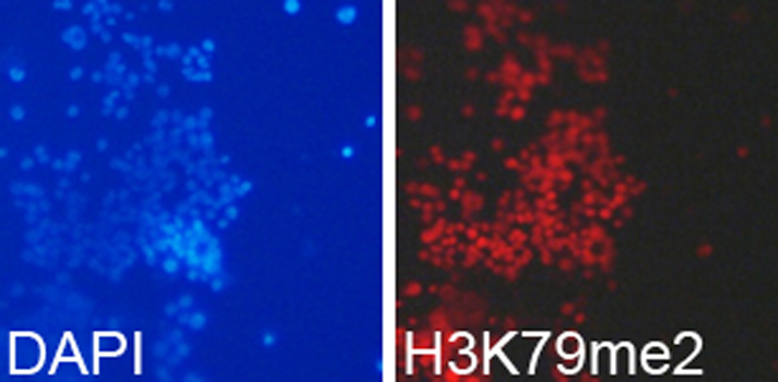
Immunofluorescence analysis of 293T cell using H3K79me2 antibody. Blue: DAPI for nuclear staining.
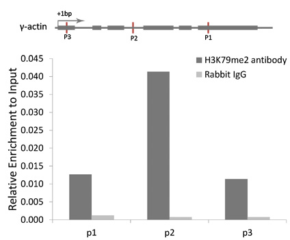
Chromatin Immunoprecipitation analysis of γ-actin gene from 293 cell line, using DiMethyl-Histone H3-K79 antibody and rabbit IgG. P1, P2 and P3 were probes located on γ-actin gene as the schematic diagram illustrated. The amount of immunoprecipitated DNA was checked by quantitative PCR. Histogram was constructed by the ratios of the immunoprecipitated DNA to the input.
Histones are basic nuclear proteins that are responsible for the nucleosome structure of the chromosomal fiber in eukaryotes. Nucleosomes consist of approximately 146 bp of DNA wrapped around a histone octamer composed of pairs of each of the four core histones (H2A, H2B, H3, and H4). The chromatin fiber is further compacted through the interaction of a linker histone, H1, with the DNA between the nucleosomes to form higher order chromatin structures. This gene is intronless and encodes a member of the histone H3 family. Transcripts from this gene lack polyA tails; instead, they contain a palindromic termination element. This gene is located separately from the other H3 genes that are in the histone gene cluster on chromosome 6p22-p21.3.
N/A


Copyrights © 2014 NEO Group 245 First Street, 18th Floor, Cambridge MA 02142 888.733.6849


