Accelerate Your Biomolecule
and Bioassay Research
and Bioassay Research
sales@neoscientific.com
+1-888.733.6849
+1-617.299.7367 (Int’l)
+1-617.299.7367 (Int’l)
+1-888.733.6849
+1-617.299.7367 (Int’l)
+1-617.299.7367 (Int’l)
For quotations, please use our online quotation form, and you may also contact us by
sales@neoscientific.com
+1-888.733.6849
+1-617.299.7367 (Int’l)
+1-888.733.6849
+1-617.299.7367 (Int’l)
| Reactivity | Human Mouse Rat |
| Tested applications | WB IHC IF |
| Recommended Dilution | WB 1:500 - 1:2000 IHC 1:50 - 1:100 IF 1:50 - 1:200 |
| Calculated MW | 38kDa |
| Observed MW | Refer to Figures |
| Immunogen | Recombinant protein of human FAS |
| Storage Buffer | Store at -20℃. Avoid freeze / thaw cycles. Buffer: PBS with 0.02% sodium azide, 50% glycerol, pH7.3. |
| Concentration | bi |
| Synonym | FAS;ALPS1A;APO-1;APT1;CD95;FAS1;FASTM;TNFRSF6 ; |

Western blot analysis of extracts of various cells, using FAS antibody.
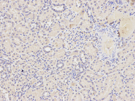
Immunohistochemistry of paraffin-embedded human kidney using FAS antibody at dilution of 1:400 (200x lens).
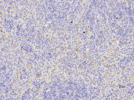
Immunohistochemistry of paraffin-embedded rat spleen using FAS antibody at dilution of 1:400 (200x lens).
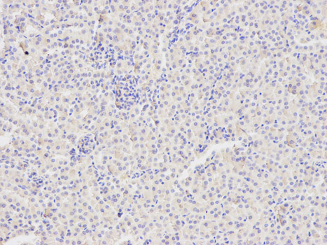
Immunohistochemistry of paraffin-embedded mouse kidney using FAS antibody at dilution of 1:400 (200x lens).
Immunohistochemistry of paraffin-embedded human prostate using FAS antibody.
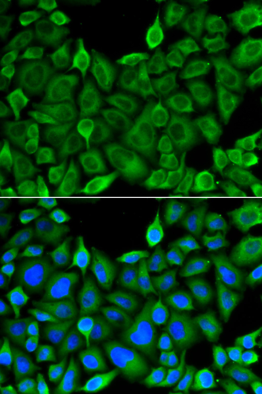
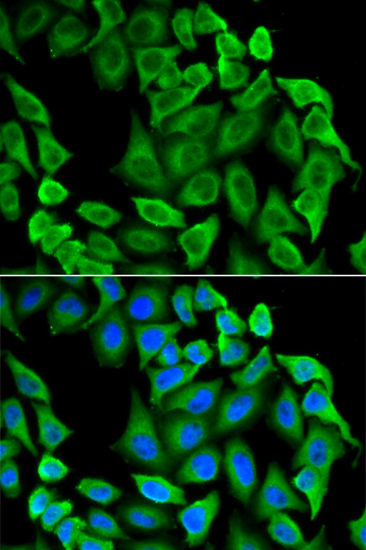
Immunofluorescence analysis of A549 cell using FAS antibody. Blue: DAPI for nuclear staining.

Immunofluorescence analysis of HeLa cell using FAS antibody. Blue: DAPI for nuclear staining.
Association of the receptor Fas with its ligand FasL triggers an apoptotic pathway that plays an important role in immune regulation, development, and progression of cancers (1,2). Loss of function mutation in either Fas (lpr mice) or FasL (gld mice) leads to lymphadenopathy and splenomegaly as a result of decreased apoptosis in CD4-CD8- T lymphocytes (3,4). FasL (CD95L, Apo-1L) is a type II transmembrane protein of 280 amino acids (runs at approximately 40 kDa upon glycosylation) that belongs to the TNF family, which also includes TNF-α, TRAIL, and TWEAK. Binding of FasL to its receptor triggers the formation of a death-inducing signaling complex (DISC) involving the recruitment of the adaptor protein FADD and caspase-8 (5). Activation of caspase-8 from this complex initiates a caspase cascade resulting in the activation of caspase-3 and subsequent cleavage of proteins leading to apoptosis. Unlike Fas, which is constitutively expressed by various cell types, FasL is predominantly expressed on activated T lymphocytes, NK cells, and at immune privileged sites (6). FasL is also expressed in several tumor types as a mechanism to evade immune surveillance (7). Similar to other members of the TNF family, FasL can be cleaved by metalloproteinases producing a 26 kDa trimeric soluble form (8,9).
N/A


Copyrights © 2014 NEO Group 245 First Street, 18th Floor, Cambridge MA 02142 888.733.6849


