Accelerate Your Biomolecule
and Bioassay Research
and Bioassay Research
sales@neoscientific.com
+1-888.733.6849
+1-617.299.7367 (Int’l)
+1-617.299.7367 (Int’l)
+1-888.733.6849
+1-617.299.7367 (Int’l)
+1-617.299.7367 (Int’l)
For quotations, please use our online quotation form, and you may also contact us by
sales@neoscientific.com
+1-888.733.6849
+1-617.299.7367 (Int’l)
+1-888.733.6849
+1-617.299.7367 (Int’l)
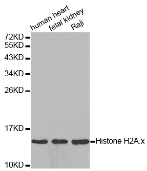
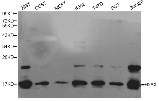
| Reactivity | Human Mouse Rat |
| Tested applications | WB IHC ICC |
| Recommended Dilution | WB 1:500 - 1:2000 IHC 1:50 - 1:200 ICC 1:50 - 1:200 |
| Calculated MW | 17kDa |
| Observed MW | Refer to Figures |
| Immunogen | Recombinant Protein of human H2AFX |
| Storage Buffer | Store at -20℃. Avoid freeze / thaw cycles. Buffer: PBS with 0.02% sodium azide, 50% glycerol, pH7.3. |
| Synonym | H2AFX;H2A.X ;H2A/X;H2AX;Histone H2A.x ; |
Histone H2A.X is a variant histone that represents approximately 10% of the total H2A histone proteins in normal human fibroblasts (1). H2A.X is required for checkpoint-mediated cell cycle arrest and DNA repair following double-stranded DNA breaks (1). DNA damage, caused by ionizing radiation, UV-light, or radiomimetic agents, results in rapid phosphorylation of H2A.X at Ser139 by PI3K-like kinases, including ATM, ATR, and DNA-PK (2,3). Within minutes following DNA damage, H2A.X is phosphorylated at Ser139 at sites of DNA damage (4). This very early event in the DNA-damage response is required for recruitment of a multitude of DNA-damage response proteins, including MDC1, NBS1, RAD50, MRE11, 53BP1, and BRCA1 (1). In addition to its role in DNA-damage repair, H2A.X is required for DNA fragmentation during apoptosis and is phosphorylated by various kinases in response to apoptotic signals. H2A.X is phosphorylated at Ser139 by DNA-PK in response to cell death receptor activation, c-Jun N-terminal Kinase (JNK1) in response to UV-A irradiation, and p38 MAPK in response to serum starvation (5-8). H2A.X is constitutively phosphorylated on Tyr142 in undamaged cells by WSTF (Williams-Beuren syndrome transcription factor) (9,10). Upon DNA damage, and concurrent with phosphorylation of Ser139, Tyr142 is dephosphorylated at sites of DNA damage by recruited EYA1 and EYA3 phosphatases (9). While phosphorylation at Ser139 facilitates the recruitment of DNA repair proteins and apoptotic proteins to sites of DNA damage, phosphorylation at Tyr142 appears to determine which set of proteins are recruited. Phosphorylation of H2A.X at Tyr142 inhibits the recruitment of DNA repair proteins and promotes binding of pro-apoptotic factors such as JNK1 (9). Mouse embryonic fibroblasts expressing only mutant H2A.X Y142F, which favors recruitment of DNA repair proteins over apoptotic proteins, show a reduced apoptotic response to ionizing radiation (9). Thus, it appears that the balance of H2A.X Tyr142 phosphorylation and dephosphorylation provides a switch mechanism to determine cell fate after DNA damage.
N/A


Copyrights © 2014 NEO Group 245 First Street, 18th Floor, Cambridge MA 02142 888.733.6849