Accelerate Your Biomolecule
and Bioassay Research
and Bioassay Research
sales@neoscientific.com
+1-888.733.6849
+1-617.299.7367 (Int’l)
+1-617.299.7367 (Int’l)
+1-888.733.6849
+1-617.299.7367 (Int’l)
+1-617.299.7367 (Int’l)
For quotations, please use our online quotation form, and you may also contact us by
sales@neoscientific.com
+1-888.733.6849
+1-617.299.7367 (Int’l)
+1-888.733.6849
+1-617.299.7367 (Int’l)
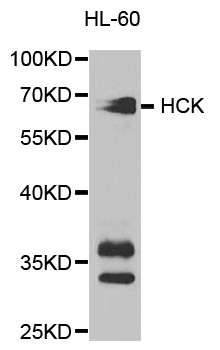
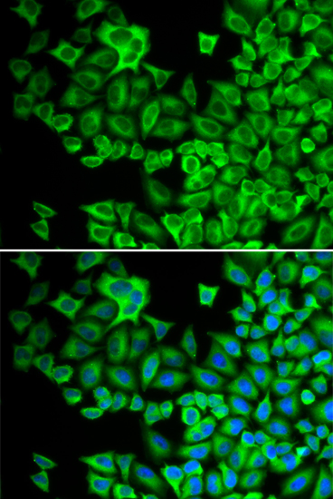
| Reactivity | Human Mouse Rat |
| Tested applications | WB IHC IF |
| Recommended Dilution | WB 1:500 - 1:2000 IHC 1:50 - 1:200 IF 1:10 - 1:100 |
| Calculated MW | 57kDa |
| Observed MW | Refer to Figures |
| Immunogen | Recombinant protein of human HCK |
| Storage Buffer | Store at -20℃. Avoid freeze / thaw cycles. Buffer: PBS with 0.02% sodium azide, 50% glycerol, pH7.3. |
| Synonym | JTK9 |
Hck (hemopoietic cell kinase) is a protein tyrosine kinase of the Src family prominently expressed in the lymphoid and myeloid lineages of hemopoiesis (1). It participates in transducing a variety of extracellular signals, which ultimately affect cellular processes including proliferation, differentiation and migration.The well-defined modular structure of Hck comprises a relatively divergent, NH2-terminal "unique" domain, which is subject to post-translational lipid modifications thereby targeting Hck to the plasma membrane. Src homology 3 (SH3) and 2 (SH2) domains, and a tyrosine kinase catalytic domain follow the "unique" domain. The catalytic activity of Hck is regulated, both positively and negatively, by tyrosine phosphorylation of highly conserved tyrosine (Y) residues. Phosphorylation of a single conserved Tyr499 residue in the COOH terminus of Hck by the protein kinase Csk renders Hck inactive as a result of an intramolecular interaction between the phosphorylated tyrosine (pY) residue and its own SH2 domain. Disruption of this interaction, either as a result of dephosphorylation, or substitution of the COOH-terminal regulatory Y residue with phenylalanine (F; e.g., HckY499F), or COOH-terminal truncation mutations as observed in the virally transduced v-Src oncoprotein, results in constitutive activation of Hck. In contrast to phosphorylation of the COOH-terminal regulatory tyrosine residue, autophosphorylation of a tyrosine residue (Tyr388) within the kinase domain of Hck acts to positively regulate its catalytic activity. Thus, activation of Hck requires both disruption of the COOH-terminal regulatory tyrosine-SH2 domain interaction and autophosphorylation of the regulatory tyrosine residue within the kinase domain ( 2, 3). The dysfunction or dysregulation of Hck may contribute to the pathogenesis of some human leukemias (4).
N/A


Copyrights © 2014 NEO Group 245 First Street, 18th Floor, Cambridge MA 02142 888.733.6849