Accelerate Your Biomolecule
and Bioassay Research
and Bioassay Research
sales@neoscientific.com
+1-888.733.6849
+1-617.299.7367 (Int’l)
+1-617.299.7367 (Int’l)
+1-888.733.6849
+1-617.299.7367 (Int’l)
+1-617.299.7367 (Int’l)
For quotations, please use our online quotation form, and you may also contact us by
sales@neoscientific.com
+1-888.733.6849
+1-617.299.7367 (Int’l)
+1-888.733.6849
+1-617.299.7367 (Int’l)
| Reactivity | Human Mouse Rat |
| Tested applications | WB IHC IF |
| Recommended Dilution | WB 1:500 - 1:2000 IHC 1:50 - 1:200 IF 1:50 - 1:200 |
| Calculated MW | 113kDa |
| Observed MW | Refer to Figures |
| Immunogen | Recombinant protein of human LIG3 |
| Storage Buffer | Store at -20℃. Avoid freeze / thaw cycles. Buffer: PBS with 0.02% sodium azide, 50% glycerol, pH7.3. |
| Concentration | e |
| Synonym | LIG2; LIG3; |
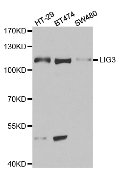
Western blot analysis of extracts of various cell lines, using LIG3 antibody(1:1000).
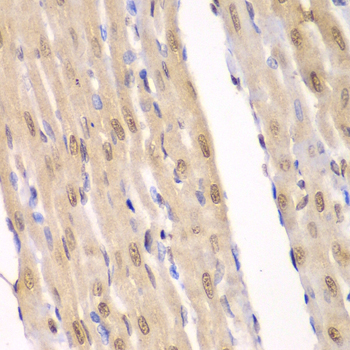
Immunohistochemistry of paraffin-embedded rat heart using LIG3 antibody at dilution of 1:100 (400x lens).
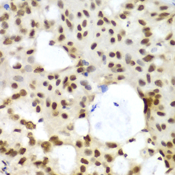
Immunohistochemistry of paraffin-embedded human oophoroma using LIG3 antibody at dilution of 1:100 (400x lens).
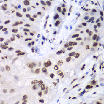
Immunohistochemistry of paraffin-embedded human esophageal cancer using LIG3 antibody at dilution of 1:100 (400x lens).
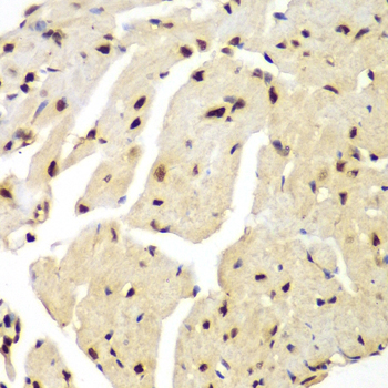
Immunohistochemistry of paraffin-embedded mouse heart using LIG3 antibody at dilution of 1:100 (400x lens).
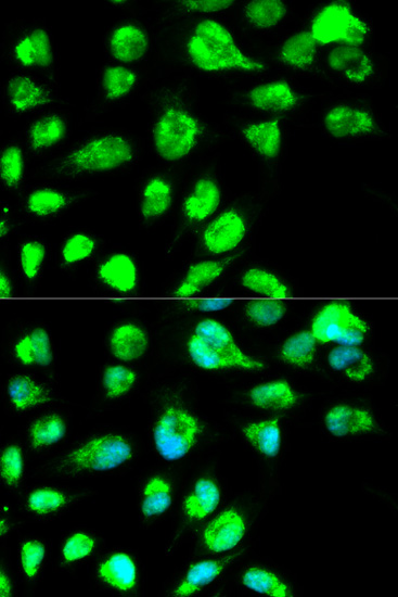
Immunofluorescence analysis of MCF7 cell using LIG3 antibody. Blue: DAPI for nuclear staining.
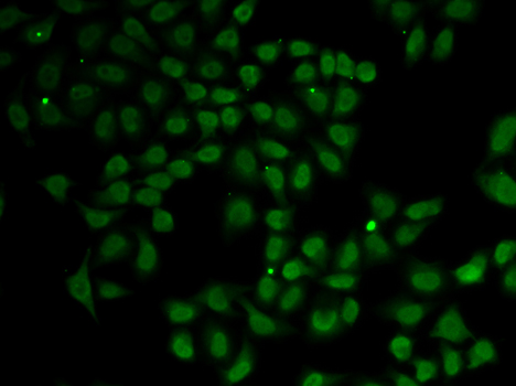
Immunofluorescence analysis of A549 cell using LIG3 antibody.
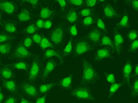
Immunofluorescence analysis of HeLa cell using LIG3 antibody.
This gene is a member of the DNA ligase family. Each member of this family encodes a protein that catalyzes the joining of DNA ends but they each have a distinct role in DNA metabolism. The protein encoded by this gene is involved in excision repair and is located in both the mitochondria and nucleus, with translation initiation from the upstream start codon allowing for transport to the mitochondria and translation initiation from a downstream start codon allowing for transport to the nucleus. Additionally, alternate transcriptional splice variants, encoding different isoforms, have been characterized. [provided by RefSeq, Jul 2008]
N/A


Copyrights © 2014 NEO Group 245 First Street, 18th Floor, Cambridge MA 02142 888.733.6849


