Accelerate Your Biomolecule
and Bioassay Research
and Bioassay Research
sales@neoscientific.com
+1-888.733.6849
+1-617.299.7367 (Int’l)
+1-617.299.7367 (Int’l)
+1-888.733.6849
+1-617.299.7367 (Int’l)
+1-617.299.7367 (Int’l)
For quotations, please use our online quotation form, and you may also contact us by
sales@neoscientific.com
+1-888.733.6849
+1-617.299.7367 (Int’l)
+1-888.733.6849
+1-617.299.7367 (Int’l)
| Reactivity | Human Mouse |
| Tested applications | WB IHC |
| Recommended Dilution | WB 1:500 - 1:2000 IHC 1:100 - 1:200 |
| Calculated MW | 37kDa |
| Observed MW | Refer to Figures |
| Immunogen | A synthetic peptide of human MCL1 |
| Storage Buffer | Store at -20℃. Avoid freeze / thaw cycles. Buffer: PBS with 0.02% sodium azide, 50% glycerol, pH7.3. |
| Concentration | bo |
| Synonym | MCL1;BCL2L3;EAT;MCL1L;MCL1S;MGC104264;MGC1839;Mcl-1;TM ; |
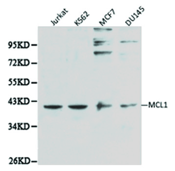
Western blot analysis of extracts of various cell lines, using MCL1 antibody.

Immunohistochemistry of paraffin-embedded mouse kidney using MCL1 antibody at dilution of 1:100 (x40 lens).

Immunohistochemistry of paraffin-embedded human lung cancer using MCL1 antibody at dilution of 1:100 (x40 lens).

Immunohistochemistry of paraffin-embedded human prostate using MCL1 antibody at dilution of 1:100 (x40 lens).

Immunohistochemistry of paraffin-embedded human stomach using MCL1 antibody at dilution of 1:100 (x40 lens).

Immunohistochemistry of paraffin-embedded human gastric cancer using MCL1 antibody at dilution of 1:100 (x40 lens).

Immunohistochemistry of paraffin-embedded mouse liver using MCL1 antibody at dilution of 1:100 (x40 lens).

Immunohistochemistry of paraffin-embedded mouse brain using MCL1 antibody at dilution of 1:100 (x40 lens).
Mcl-1 is an anti-apoptotic member of the Bcl-2 family originally isolated from the ML-1 human myeloid leukemia cell line during phorbol ester-induced differentiation along the monocyte/macrophage pathway (1). Similar to other Bcl-2 family members, Mcl-1 localizes to the mitochondria (2), interacts with and antagonizes pro-apoptotic Bcl-2 family members (3), and inhibits apoptosis induced by a number of cytotoxic stimuli (4). Mcl-1 differs from its other family members in its regulation at both the transcriptional and post-translational level. First, Mcl-1 has an extended amino-terminal PEST region, which is responsible for its relatively short half-life (1,2). Second, unlike other family members, Mcl-1 is rapidly transcribed via a PI3K/Akt dependent pathway, resulting in its increased expression during myeloid differentiation and cytokine stimulation (1,5-7). Mcl-1 is phosphorylated in response to treatment with phorbol ester, microtubule-damaging agents, oxidative stress, and cytokine withdrawal (8-11). Phosphorylation at Thr163, the conserved MAP kinase/ERK site located within the PEST region, slows Mcl-1 protein turnover (10) but may prime the GSK-3 mediated phosphorylation at Ser159 that leads to Mcl-1 destabilization (11). Mcl-1 deficiency in mice results in peri-implantation lethality (12). In addition, conditional disruption of the corresponding mcl-1 gene shows that Mcl-1 plays an important role in early lymphoid development and in the maintenance of mature lymphocytes (13).
N/A


Copyrights © 2014 NEO Group 245 First Street, 18th Floor, Cambridge MA 02142 888.733.6849


