Accelerate Your Biomolecule
and Bioassay Research
and Bioassay Research
sales@neoscientific.com
+1-888.733.6849
+1-617.299.7367 (Int’l)
+1-617.299.7367 (Int’l)
+1-888.733.6849
+1-617.299.7367 (Int’l)
+1-617.299.7367 (Int’l)
For quotations, please use our online quotation form, and you may also contact us by
sales@neoscientific.com
+1-888.733.6849
+1-617.299.7367 (Int’l)
+1-888.733.6849
+1-617.299.7367 (Int’l)
| Reactivity | Human Mouse Rat |
| Tested applications | WB IHC IF |
| Recommended Dilution | WB 1:500 - 1:2000 IHC 1:50 - 1:200 IF 1:50 - 1:200 |
| Calculated MW | 78kDa;101kDa |
| Observed MW | Refer to Figures |
| Immunogen | Recombinant protein of human NFATC1 |
| Storage Buffer | Store at -20℃. Avoid freeze / thaw cycles. Buffer: PBS with 0.02% sodium azide, 50% glycerol, pH7.3. |
| Concentration | k |
| Synonym | MGC138448; NF-ATC; NFAT2; NFATc; |
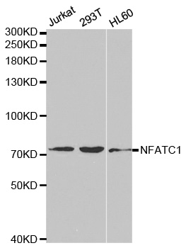
Western blot analysis of extracts of various cell lines, using NFATC1 antibody.
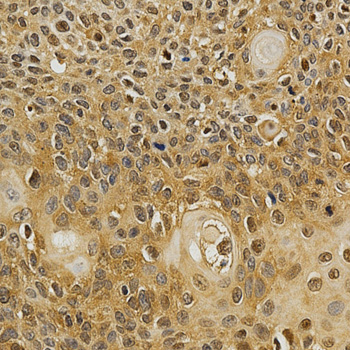
Immunohistochemistry of paraffin-embedded human esophageal cancer using NFATC1 antibody at dilution of 1:200 (x400 lens).
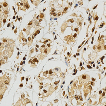
Immunohistochemistry of paraffin-embedded human stomach using NFATC1 antibody at dilution of 1:200 (x400 lens).

Immunohistochemistry of paraffin-embedded Human esophageal using NFATC1 antibody at dilution of 1:100 (x400 lens).
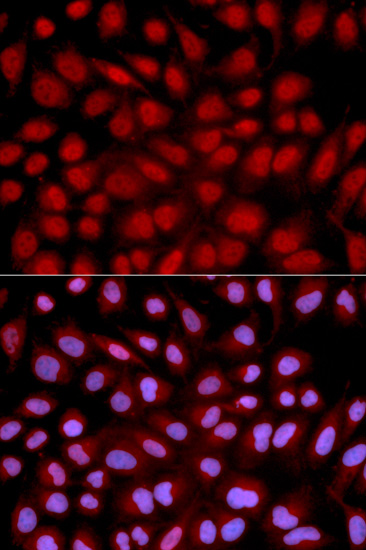
Immunofluorescence analysis of U2OS cell using NFATC1 antibody. Blue: DAPI for nuclear staining.
The NFAT (nuclear factor of activated T cells) family of proteins consists of NFAT1 (NFATc2 or NFATp), NFAT2 (NFATc1 or NFATc), NFAT3 (NFATc4), and NFAT4 (NFATc3 or NFATx). All members of this family are transcription factors with a Rel homology domain and regulate gene transcription in concert with AP-1 (Jun/Fos) to orchestrate an effective immune response (1,2). NFAT proteins are predominantly expressed in cells of the immune system, but are also expressed in skeletal muscle, keratinocytes, and adipocytes, regulating cell differentiation programs in these cells (3). In resting cells, NFAT proteins are heavily phosphorylated and localized in the cytoplasm. Increased intracellular calcium concentrations activate the calcium/calmodulin-dependent serine phosphatase calcineurin, which dephosphorylates NFAT proteins, resulting in their subsequent translocation to the nucleus (2). Termination of NFAT signaling occurs upon declining calcium concentrations and phosphorylation of NFAT by kinases such as GSK-3 or CK1 (3,4). Cyclosporin A and FK506 are immunosuppressive drugs that inhibit calcineurin and thus retain NFAT proteins in the cytoplasm (5).
N/A


Copyrights © 2014 NEO Group 245 First Street, 18th Floor, Cambridge MA 02142 888.733.6849


