Accelerate Your Biomolecule
and Bioassay Research
and Bioassay Research
sales@neoscientific.com
+1-888.733.6849
+1-617.299.7367 (Int’l)
+1-617.299.7367 (Int’l)
+1-888.733.6849
+1-617.299.7367 (Int’l)
+1-617.299.7367 (Int’l)
For quotations, please use our online quotation form, and you may also contact us by
sales@neoscientific.com
+1-888.733.6849
+1-617.299.7367 (Int’l)
+1-888.733.6849
+1-617.299.7367 (Int’l)
| Reactivity | Human Mouse Rat |
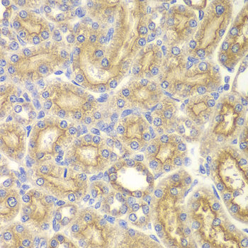
| Tested applications | WB IHC ICC IP |
| Recommended Dilution | WB 1:500 - 1:2000 IHC 1:50 - 1:200 ICC 1:50 - 1:200 IP 1:20 - 1:100 |
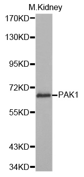
| Calculated MW | 62kDa |
| Observed MW | Refer to Figures |
| Immunogen | Recombinant protein of human PAK1 |
| Storage Buffer | Store at -20℃. Avoid freeze / thaw cycles. Buffer: PBS with 0.02% sodium azide, 50% glycerol, pH7.3. |
| Concentration | bfqr |
| Synonym | MGC130000; MGC130001; PAKalpha; |
The p21-activated kinase (PAK) family of serine/threonine kinases is engaged in multiple cellular processes, including cytoskeletal reorganization, MAPK signaling, apoptotic signaling, control of phagocyte NADPH oxidase, and growth factor-induced neurite outgrowth (1,2). Several mechanisms that induce PAK activity have been reported. Binding of Rac/Cdc42 to the CRIB (or PBD) domain near the amino terminus of PAK causes autophosphorylation and conformational changes in PAK (1). Phosphorylation of PAK1 at Thr423 by PDK induces activation of PAK1 (3). Several autophosphorylation sites have been identified, including Ser199 and Ser204 of PAK1 and Ser192 and Ser197 of PAK2 (4,5). Because the autophosphorylation sites are located in the amino-terminal inhibitory domain, it has been hypothesized that modification in this region prevents the kinase from reverting to an inactive conformation (6). Research indicates that phosphorylation at Ser144 of PAK1 or Ser139 of PAK3 (located in the kinase inhibitory domain) affects kinase activity (7). Phosphorylation at Ser21 of PAK1 or Ser20 of PAK2 regulates binding with the adaptor protein Nck (8). PAK4, PAK5, and PAK6 have lower sequence similarity with PAK1-3 in the amino-terminal regulatory region (9). Phosphorylation at Ser474 of PAK4, a site analogous to Thr423 of PAK1, may play a pivotal role in regulating the activity and function of PAK4 (10).
N/A


Copyrights © 2014 NEO Group 245 First Street, 18th Floor, Cambridge MA 02142 888.733.6849