Accelerate Your Biomolecule
and Bioassay Research
and Bioassay Research
sales@neoscientific.com
+1-888.733.6849
+1-617.299.7367 (Int’l)
+1-617.299.7367 (Int’l)
+1-888.733.6849
+1-617.299.7367 (Int’l)
+1-617.299.7367 (Int’l)
For quotations, please use our online quotation form, and you may also contact us by
sales@neoscientific.com
+1-888.733.6849
+1-617.299.7367 (Int’l)
+1-888.733.6849
+1-617.299.7367 (Int’l)
| Reactivity | Human Mouse Rat |
| Tested applications | WB IHC ICC IF IP |
| Recommended Dilution | WB 1:500 - 1:2000 IHC 1:50 - 1:100 ICC 1:50 - 1:200 IF 1:50 - 1:200 IP 1:20 - 1:100 |
| Calculated MW | 51kDa |
| Observed MW | Refer to Figures |
| Immunogen | A synthetic peptide of human RUNX1 |
| Storage Buffer | Store at -20℃. Avoid freeze / thaw cycles. Buffer: PBS with 0.02% sodium azide, 50% glycerol, pH7.3. |
| Concentration | akn |
| Synonym | AML1; AML1-EVI-1; AMLCR1; CBFA2; EVI-1; PEBP2Ab; |
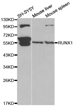
Western blot analysis of extracts of various cell lines, using RUNX1 antibody.
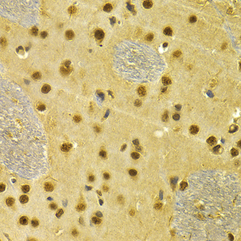
Immunohistochemistry of paraffin-embedded rat brain using RUNX1 antibody at dilution of 1:100 (x40 lens).
Immunohistochemistry of paraffin-embedded rat kidney using RUNX1 antibody at dilution of 1:100 (x40 lens).
Immunohistochemistry of paraffin-embedded mouse kidney using RUNX1 antibody at dilution of 1:100 (x40 lens).
Immunohistochemistry of paraffin-embedded human stomach using RUNX1 antibody at dilution of 1:100 (x40 lens).

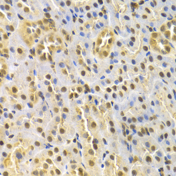
Immunohistochemistry of paraffin-embedded rat liver using RUNX1 antibody at dilution of 1:100 (x40 lens).

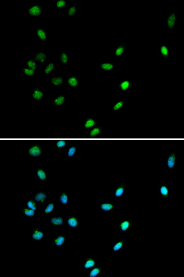
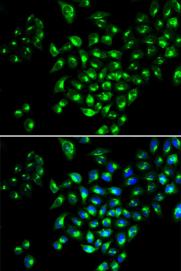
Immunofluorescence analysis of A549 cell using RUNX1 antibody. Blue: DAPI for nuclear staining.

Immunofluorescence analysis of MCF-7 cell using RUNX1 antibody. Blue: DAPI for nuclear staining.
AML1 (also known as Runx1, CBFA2, and PEBP2αB) is a member of the core binding factor (CBF) family of transcription factors (1,2). It is required for normal development of all hematopoietic lineages (3-5). AML1 forms a heterodimeric DNA binding complex with its partner protein CBFβ and regulates the expression of cellular genes by binding to promoter and enhancer elements. AML1 is commonly translocated in hematopoietic cancers: chromosomal translocations include t(8;21) AML1-ETO, t(12;21) TEL-AML, and t(8;21) AML-M2 (6). Phosphorylation of AML1 on several potential serine and threonine sites, including Ser249, is thought to occur in an Erk-dependent manner (7,8).
N/A


Copyrights © 2014 NEO Group 245 First Street, 18th Floor, Cambridge MA 02142 888.733.6849


