Accelerate Your Biomolecule
and Bioassay Research
and Bioassay Research
sales@neoscientific.com
+1-888.733.6849
+1-617.299.7367 (Int’l)
+1-617.299.7367 (Int’l)
+1-888.733.6849
+1-617.299.7367 (Int’l)
+1-617.299.7367 (Int’l)
For quotations, please use our online quotation form, and you may also contact us by
sales@neoscientific.com
+1-888.733.6849
+1-617.299.7367 (Int’l)
+1-888.733.6849
+1-617.299.7367 (Int’l)
| Reactivity | Human Mouse |
| Tested applications | WB IHC ICC IP |
| Recommended Dilution | WB 1:500 - 1:2000 IHC 1:50 - 1:200 ICC 1:50 - 1:200 IP 1:20 - 1:50 |
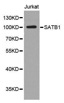
| Calculated MW | 100kDa |
| Observed MW | Refer to Figures |
| Immunogen | A synthetic peptide of human SATB1 |
| Storage Buffer | Store at -20℃. Avoid freeze / thaw cycles. Buffer: PBS with 0.02% sodium azide, 50% glycerol, pH7.3. |
| Synonym | SATB1;DNA-binding protein SATB1; |
Special AT-rich binding protein 1 (SATB1) functions as both a global chromatin organizer and a gene-specific transcription factor (1). SATB1 cooperates with promyelocytic leukemia protein (PML) to regulate global chromatin architecture by organizing chromatin into distinct loops via periodic anchoring of matrix attachment regions (MARs) in DNA to the nuclear matrix (1-3). In addition, SATB1 recruits multiple chromatin-remodeling proteins that contribute to specific gene activation and repression, including the chromatin remodeling enzymes ACF and ISWI, the histone deacetylase HDAC1, and the histone acetyltransferases PCAF and p300/CBP (4-6). Phosphorylation of SATB1 on Ser185 by protein kinase C regulates its interaction with HDAC1 and PCAF. While unphosphorylated SATB1 binds to PCAF, phosphorylated SATB1 preferentially binds to HDAC1 (6). Acetylation of SATB1 on Lys136 by PCAF impairs its DNA binding activity, thereby removing SATB1 from gene promoters (6). SATB1 is expressed predominantly in thymocytes and is involved in gene regulation during T cell activation (1). SATB1 is also expressed in metastatic breast cancer cells and is a potential prognostic marker and therapeutic target for metastatic breast cancer (7). In a mouse model system, RNAi-mediated knockdown of SATB1 reversed tumorigenesis by inhibiting tumor growth and metastasis, while ectopic expression of SATB1 in non-metastatic breast cancer cells produced invasive tumors.Phospho-SATB1 (Ser47) Antibody is directed at a site that was identified at Cell Signaling Technology (CST) using PhosphoScan®, CST's LC-MS/MS platform for modification site discovery. Phosphorylation at Ser47 was discovered using an Akt substrate antibody. The function of this phosphorylation event is not known. Please visit PhosphoSitePlus™, CST's modification site knowledgebase, at www.phosphosite.org for more information.
N/A


Copyrights © 2014 NEO Group 245 First Street, 18th Floor, Cambridge MA 02142 888.733.6849