Accelerate Your Biomolecule
and Bioassay Research
and Bioassay Research
sales@neoscientific.com
+1-888.733.6849
+1-617.299.7367 (Int’l)
+1-617.299.7367 (Int’l)
+1-888.733.6849
+1-617.299.7367 (Int’l)
+1-617.299.7367 (Int’l)
For quotations, please use our online quotation form, and you may also contact us by
sales@neoscientific.com
+1-888.733.6849
+1-617.299.7367 (Int’l)
+1-888.733.6849
+1-617.299.7367 (Int’l)
| Reactivity | Human Mouse Rat |
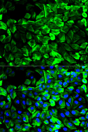
| Tested applications | WB IHC IF |
| Recommended Dilution | WB 1:500 - 1:2000 IHC 1:50 - 1:200 IF 1:50 - 1:200 |
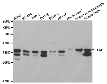
| Calculated MW | 33kDa |
| Observed MW | Refer to Figures |
| Immunogen | Recombinant protein of human TPM1 |
| Storage Buffer | Store at -20℃. Avoid freeze / thaw cycles. Buffer: PBS with 0.02% sodium azide, 50% glycerol, pH7.3. |
| Synonym | CMH3; TMSA; CMD1Y; C15orf13; HTM-alpha |
Tropomyosin-1 (TPM1) belongs to the high molecular weight members of tropomyosin family (1,2). The protein exists in an alpha-helical coiled-coil conformation and binds multiple acting monomers in a tight manner to stabilize and regulate the actin filament (3). Tropomyosins fullfill functions in muscle and non-muscle cells. In muscle cells, tropomyosins associate with the troponin complex and play a central role in the calcium-dependent regulation of striated muscle contraction in vertebrates. In non-muscle cells, tropomyosins are implicated in the formation and stabilization of cytoskeletal actin filaments to ensure normal cellular processes (1,2). Mutations of tropomysin-1 have been reported as a cause of dilated cardiac myopathies (4). Tropomyosin-1 also functions as a tumor suppressor, and many malignant tumors demonstrate downregulation of tropomyosin-1 expression (5-8). Tropomyosin-1 is phosphorylated at Ser283 through the Erk/DAPK pathway, which promotes stress fiber formation in response to oxidative stress (9-10).
N/A


Copyrights © 2014 NEO Group 245 First Street, 18th Floor, Cambridge MA 02142 888.733.6849